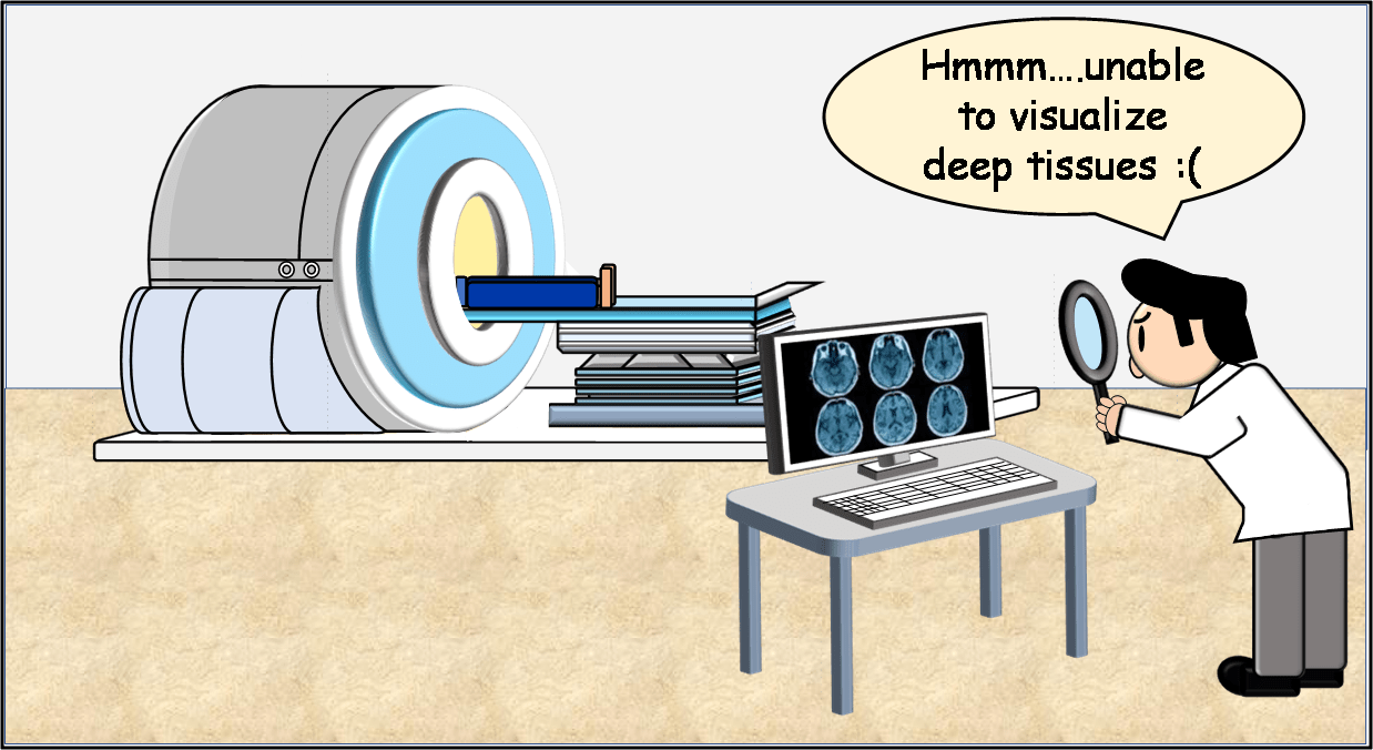
You must have heard of an MRI. Magnetic Resonance Imaging, a neuroimaging technique, uses a strong magnetic field and radio waves to get amazing pictures of our brains. Neuroimaging, or brain scanning, is used for the functional and structural imaging of the brain. Most often it is difficult to obtain clear images as we dive deeper into the brain tissues. Microscopists have been trying to obtain high-quality images of deep tissues in the brain for a long time. But they face problems in obtaining such images in a short span.
Recently, a group of researchers have developed a novel technique that can produce deep tissue images at high resolution. This process involves computational imaging to generate images at a speed about 1000 times higher than other methods.
Microscopy for brain imaging
The advances in microscopic techniques and neurotracing methods have helped us develop a better understanding of the brain structure. Most studies on brain anatomy generate a large amount of anatomical data that has to be further analyzed by computational methods. This helps to answer specific questions.
Light microscopy offers several benefits for in-vivo brain imaging. It provides a non-invasive and highly sensitive method for detecting functional changes in the brain. Light microscopy can also provide a good contrast by using both internal and external chromophores. Internal chromophores involve cytochrome, metabolites, deoxyhemoglobin, etc. These have distinct fluorescent and absorbance properties. Many biomolecules have natural optical contrast. This increases the availability of contrast agents for this technique. Also, the instruments for light imaging are less expensive than other techniques like PET or MRI.
However, optical imaging has certain limitations. The phenomenon of light scattering can limit image resolution. Thus, different variations of light microscopy are required to overcome this effect.

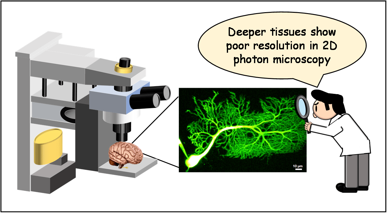
It is common to use two-photon microscopy to generate 3-D images of the brain.
The two-photon microscopy uses a high-intensity laser to cause fluorescence excitation of the specimen. The fluorescent dye has to be injected into the brain cortex to generate contrast.
The laser is focused at a single point in the sample. This is a point-by-point scanning technique as two photons are absorbed in a single focal point with the highest intensity. Laser light has a low wavelength and energy. This allows it to penetrate deeply into brain tissues without causing any damage. The technique is widespread in neurosciences. Apart from the difficulty in sample preparation, the laser cost and equipment for this process are high. Live imaging of the animal is also difficult. It is time-consuming as we have to scan each pixel one at a time. Most people prefer to build their variations of 2D photon microscopy.
The “DEEP” system
2D photon microscopy can produce good quality images. Fluorescence-derived images begin to blur as we go deeper into the brain’s tissues due to light scattering. There is an option of using wide-field imaging as well. This can illuminate an entire plane of tissue at once. This speeds up the process but gives poor resolution than point-by-point scanning.
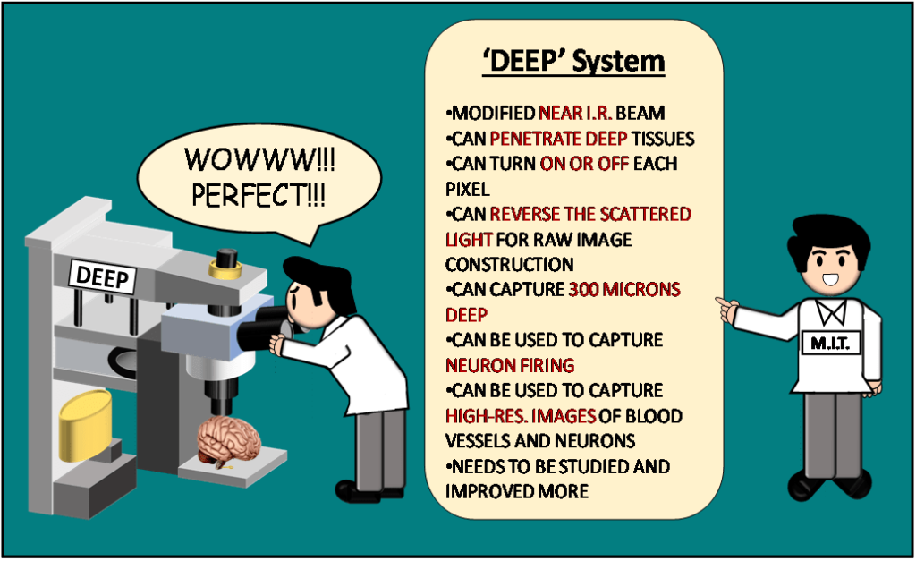
MIT researchers developed a method that could illuminate a large tissue at once while maintaining the resolution of point-by-point scanning. This technique is called “DEEP” or De-scattering with Excitation Patterning.
They modified the near-infrared laser beam so that it could penetrate deep through brain tissues. By modifying the light’s amplitude, they could turn on or off each pixel at different points of time. Laser-excited fluorescent molecules in the tissue emit signals. These signals are captured by the microscope to form an image. Some pixels remain lit up while others remain dark.
This forms a predesigned pattern that can be deduced in the light scattered by the tissue.
The computational algorithm for imaging recognizes this initial pattern and gathers information for reversing the scattered light. Researchers rebuild the raw image to reduce the scattering in the image, making the image highly resolved. The DEEP system uses structural information from millions to thousands of measurements to reconstruct the image. The research team could image up to 300 microns deep in the brain tissue. This method led to finer images than possible by other optical techniques.
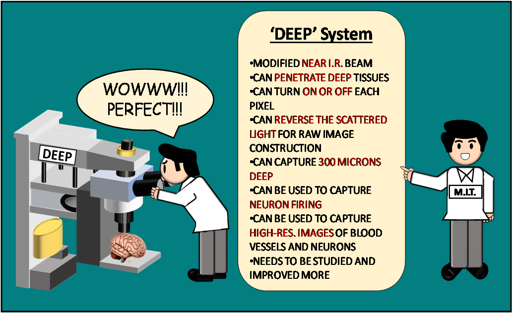
DEEP can be used to capture dynamic processes such as the firing of neurons. This might help in getting high-resolution images of blood vessels and neurons in the brain. The imaging of blood vessels of the brain could provide more information about how the flow of blood is affected by Alzheimer’s. Voltage-sensitive fluorescent dyes can be added to study neuronal activity.
DEEP system has a lot of room for improvement in the future. Even though it is in its nascent stage, this system has a lot of potential in neuroimaging.
…
Sources
- Cheng Zheng, Jong Kang Park, Murat Yildirim, Josiah R. Boivin, Yi Xue, Mriganka Sur, Peter T. C. So, Dushan N. Wadduwage.( 2021). De-scattering with Excitation Patterning enables rapid wide-field imaging through scattering media. Science Advances; 7 (28): eaay5496 DOI: 10.1126/sciadv.aay5496
- Hillman E. M. (2007). Optical brain imaging in vivo: techniques and applications from animal to man. Journal of biomedical optics, 12(5), 051402. https://doi.org/10.1117/1.2789693
- Harvard University. (2021, July 7). New imaging technique may boost research in biology, neuroscience: Scientists hope it will allow them to see inner workings of systems. ScienceDaily. Retrieved July 28, 2021 from www.sciencedaily.com/releases/2021/07/210707140642.htm
- https://scitechdaily.com/microscopy-technique-makes-high-resolution-images-of-deeper-tissue-more-quickly/
- https://courses.lumenlearning.com/boundless-psychology/chapter/brain-imaging-techniques/
Writer

Urvi Agarwal
Urvi is a biotechnology graduate. Her love for biology and writing motivated her to share interesting science stories. Currently, she is pursuing her interest in writing as a freelancer. In the future, she would like to make a career in scientific research and also continue to communicate science.
Illustrator

Jayakrishnan Nair
He has completed his Masters in Medical Biotechnology from The Maharaja Sayajirao University of Baroda and is currently an active researcher in Molecular Epidemiology at The Centre for Cancer Epidemiology, Navi Mumbai. His journey in Science and Creativity began together during his childhood days. He believes that Art and Science indeed have a beautiful world altogether in them and sailing across its spectrum inspires him to sow his thoughts deep into Science and carve out the pillars of Art through them! Being a Science enthusiast and an Artist, he wishes to devote a good share of his life to wildlife and marine conservation as he feels that the creativity of nature needs to be taken care of in the current era.